
Materials
- Lab coat
- Eye protection
- Disposable gloves
- Benchkote if necessary
- Disinfectant and paper towels
- Discard jar with disinfectant
- Bunsen burner and mat
- Microscope
- Lens tissues
- Glass slide and coverslip
- Wire loop
- Sterile water
- Plate culture of yeast
- Neutral red
- Fibre free blotting paper
 Instructions
1. Wear a lab coat, disposable gloves and use eye protection.
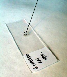
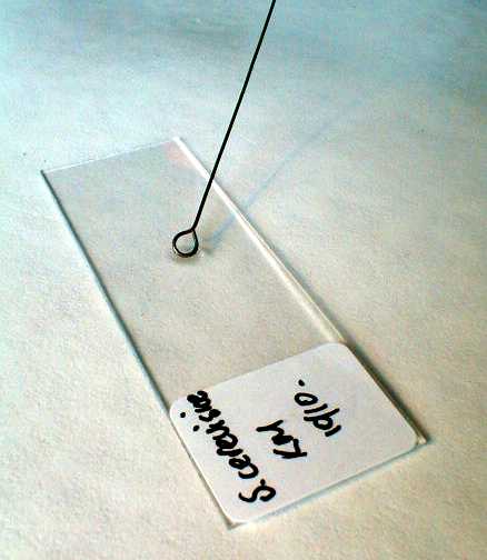
2. Clean slide and coverslip.
3. Using aseptic technique, transfer two loopfuls of sterile water to the centre of the slide
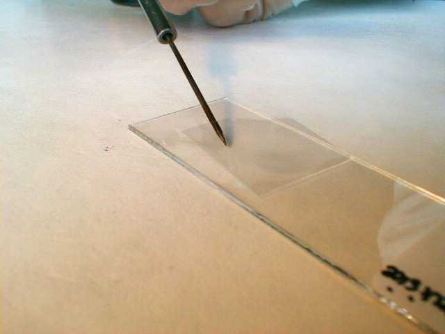
4. Using aseptic technique, transfer a small amount of yeast from a single colony into the water on the slide and mix.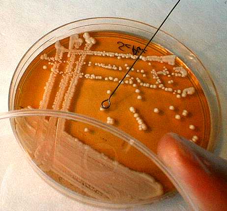
 5. Carefully lower the coverslip.
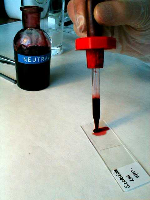 6. Using the Pasteur pipette, draw up a little neutral red.
7. Slowly release the stain along one edge of the coverslip.
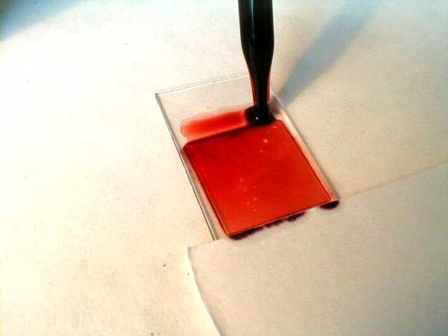 8. Place the edge of the blotting paper against the opposite edge of the coverslip to draw through the stain.
9. Observe under high power (x40) of the microscope.
10. Record the colour of the background and the colour of the cells at five minute intervals for a period of twenty minutes.
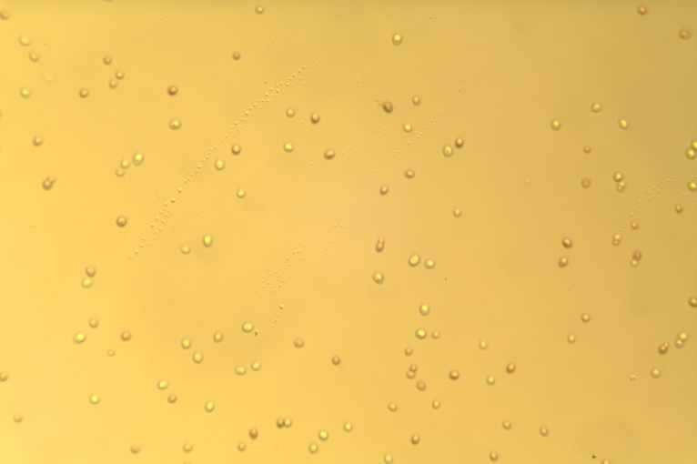
| Saccharomyces cerevisieae x160 |
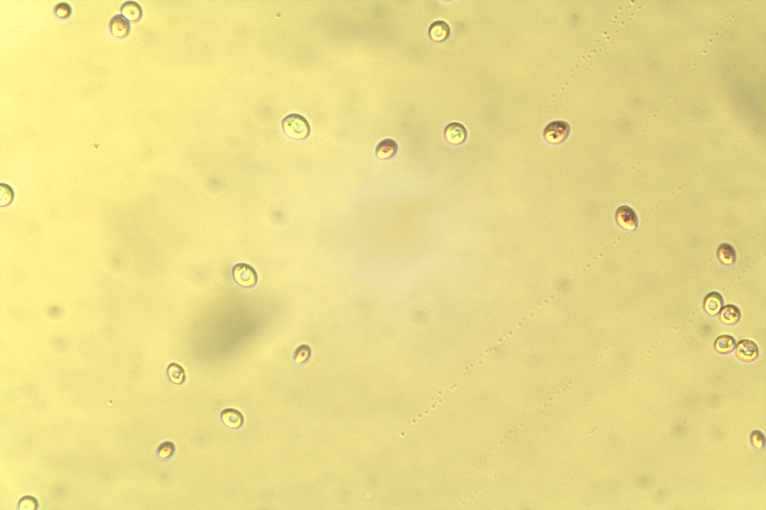
Saccharomyces cerevisieae x400 |
 |
 |

