Materials
- Lab coat
- Eye protection
- Disposable gloves
- Benchkote if necessary
- Disinfectant and paper towels
- Discard jar with disinfectant
- Fibre free blotting paper
- Labels
- Forceps
- Staining rack and dish
- Distilled water bottle
- Methylene blue stain
- Fixed smears of bacteria or yeast
Instructions
- Wear a lab coat, disposable gloves and use eye protection.
- Place the fixed smears of bacteria on a staining rack over a sink or staining tray (pie dish).
- Flood with methylene blue and leave for 2 minutes.
- Hold the slide at a 45 angle over the sink or staining tray, use the wash bottle to rinse the smear well with water.
- Blot dry between two layers of fibre free blotting paper, taking care not to rub off the cells and allow to dry in air.
- Examine under oil immersion if possible. Otherwise, under x600 (x15 eyepiece, x40 objective).
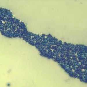
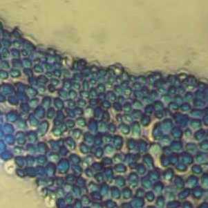
- Record shape (rod, spherical or spiral) and arrangement (clusters, chains or pairs) of the bacteria examined.
- When finished, dispose of slides into discard jar.
N.B. This method can be used for other simple stains.
Saccharomyces cervisiae
x160 x400


