
Materials
- Lab coat
- Eye protection
- Disposable plastic gloves
- Benchkote if necessary
- Disinfectant and paper towels
- Discard jar with disinfectant
- Bunsen burner and mat
- Lens tissues
- Glass slide and coverslip
- Forceps & mounted needle
- Covered beaker containing ethanol
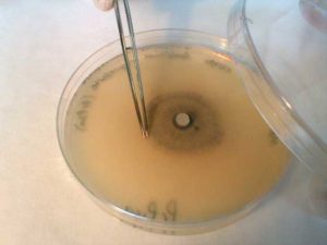
- Plate culture of mould fungus
- Lactophenol blue (note: keep lactophenol off the skin)
- Microscope
 Instructions

1. Wear a lab coat, disposable gloves and use eye protection.
2. Clean slide.
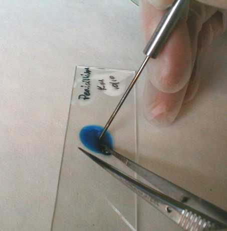
3. Place a drop of lactophenol blue in middle of slide.
4. Flame forceps with ethanol and replace lid.
5. Using aseptic technique, partially lift the lid of the Petri dish and use forceps to remove a small piece of the fungal colony.
 6. Replace lid.
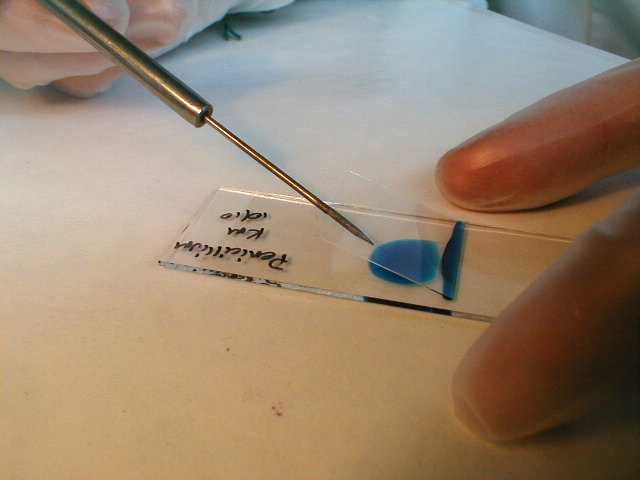
7. Place fungus in lactophenol blue and tease out well using forceps and needle.
8. Flame forceps and needle with ethanol.
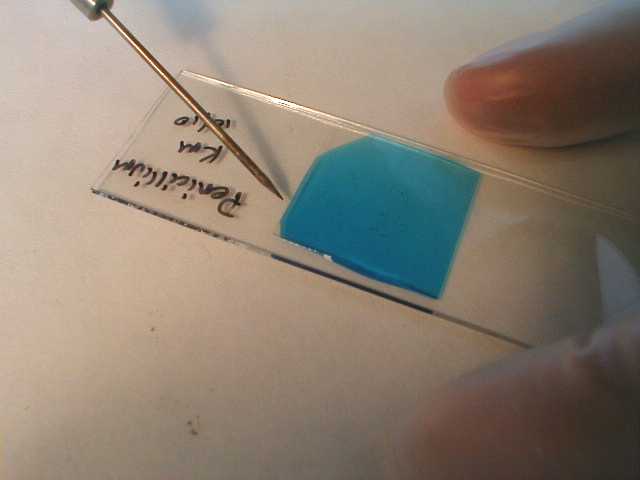
9. Using the mounted needle to support the coverslip, carefully lower it over the preparation. Take care to avoid producing bubbles.
 |
 |
10. Examine under the microscope using the x40 objective lens.
Images
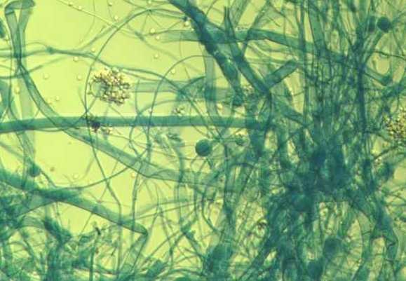
| Mucor hiemalis x160 | Mucor hiemalis x160 |
 |
 |
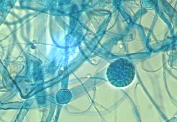
| Mucor hiemalis x400 | Mucor hiemalis x400 |
 |
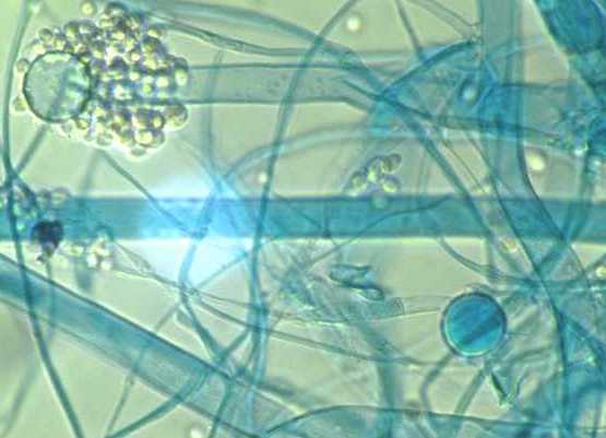 |
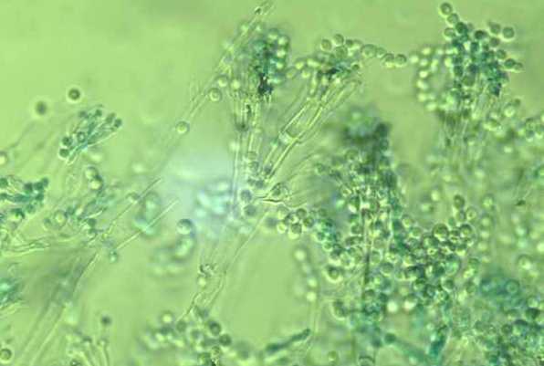
| Penicillium roquefortii x160 | |
 |
|
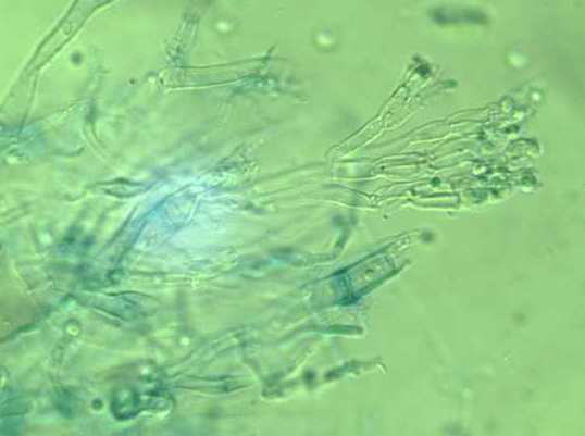
| Penicillium roquefortii x400 | Penicillium roquefortii x400 |
 |
 |

