
Materials
- Lab coat
- Eye protection
- Disposable gloves
- Benchkote if necessary
- Disinfectant and paper towels
- Discard jar with disinfectant
- Fixed smears of bacteria
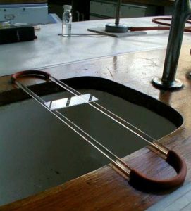
- Staining rack
- Crystal violet
- Gram’s iodine
- 95% ethanol
- Safranin
- Labels
- Forceps
- Fibre free blotting paper
- Distilled water bottle
- Bunsen burner and mat
 Instructions

1. Wear a lab coat, disposable gloves and use eye protection.
2. Lay the fixed smears of bacteria on a staining rack over a sink or staining tray (pie dish).
 3. Flood with crystal violet and leave for 1 minute.
 4. Flood with Gram’s iodine for 1 minute. (The iodine acts as a mordant i.e. a substance which increases the affinity of the bacterial cell for the dye).
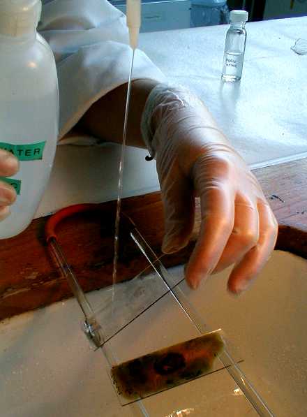
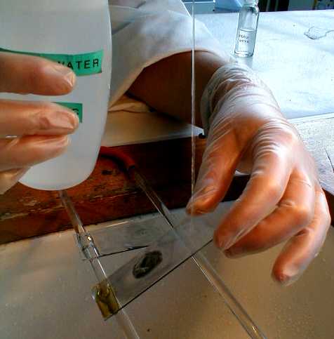
 5. Hold the slide at a 45° angle and use the wash bottle to rinse the slide well with water. Drain.
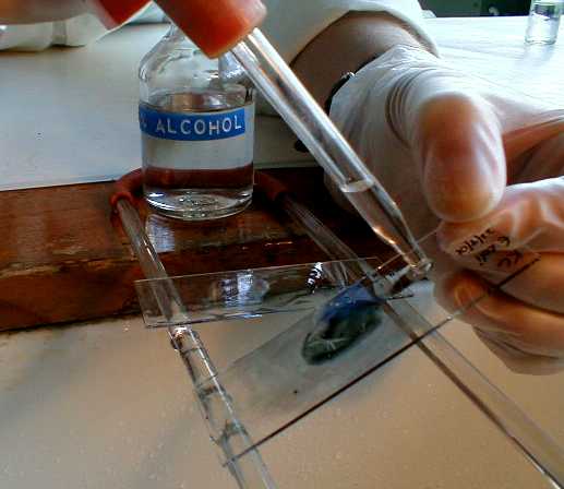
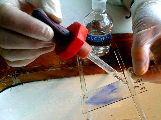 6. Hold the slide at a 45° angle over the sink or staining tray, apply the alcohol at the top of the slide with the dropper and allow the decolouriser to run down the slide over the smear, then rinse immediately with water as in step 5.
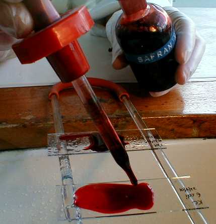 7. Flood with safranin for 1 minute.
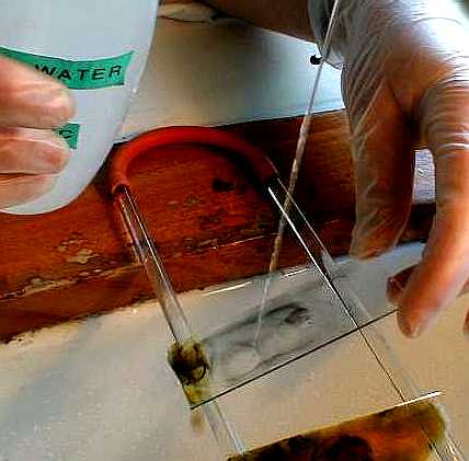
 8. Wash well with water.
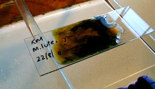
9. Blot and allow to dry in air.
 |
 |
 |
10. Examine under oil immersion if possible. Otherwise, under x600 (x40 objective, x15 eyepiece).
11. Record the Gram reaction (positive or negative), shape (rod, spherical or spiral) and arrangement (clusters, chains or pairs) of the bacteria examined.
12. When finished, dispose of slides into a discard jar.


